
X-ray is a form of electromagnetic radiation. They are waves or particles that travel though the air, similar to light or radio signals. These X-ray particles are called photons, and they are able to pass through the body and onto x-ray detectors (such as film or a detector linked to a computer monitor).
Structures that are dense (such as bone) will block most of the x-ray particles, and will appear white. Metal and contrast media (special dye used to highlight areas of the body) will also appear white. Structures containing air will be black, and muscle, fat, and fluid will appear as shades of gray.
Since Röntgen's discovery that X-rays can identify bone structures, X-rays have been developed for their use in medical imaging. Radiology is a specialized field of medicine. Radiologists employ radiography and other techniques for diagnostic imaging. This is probably the most common use of X-ray technology.
X-rays are especially useful in the detection of pathology of the skeletal system, but are also useful for detecting some disease processes in soft tissue. Some notable examples are the very common chest X-ray, which can be used to identify lung diseases such as pneumonia, lung cancer or pulmonary edema, and the abdominal X-ray, which can detect intestinal obstruction, free air (from visceral perforations) and free fluid (in ascites). X-rays may also be used to detect pathology such as gallstones (which are rarely radiopaque) or kidney stones which are often (but not always) visible. Traditional plain X-rays are less useful in the imaging of soft tissues such as the brain or muscle.
Since 2005, X-rays are listed as a carcinogen by the U.S. government. The use of X-rays as a treatment is known as radiotherapy and is largely used for the management (including palliation) of cancer; it requires higher radiation energies than for imaging alone.
X-rays are a relatively safe method of investigation and the radiation exposure is low. But in pregnant patients, the benefits of the investigation (X-ray) should be balanced with the potential hazards to the unborn fetus.
Alternative Names
Radiography
How the Test is Performed
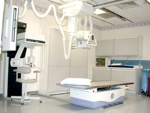
The test is performed in a hospital radiology department or in the health care provider's office by an x-ray technologist. The positioning of the patient, x-ray machine, and film depends on the type of study and area of interest. Multiple individual views may be requested.
Much like conventional photography, motion causes blurry images on radiographs, and thus, patients may be asked to hold their breath or not move during the brief exposure (about 1 second).
Preparations before the Test
Prior to the exam, the patient/client should inform her health care provider if she is pregnant, may be pregnant, or have an IUD inserted.
If abdominal studies are planned and if the patient have had a barium contrast study (such as a barium enema, upper GI series, or barium swallow) or taken medications containing bismuth (such as Pepto-Bismol) in the last 4 days, the test may be delayed until the contrast has fully passed.
All jewelry and other metallic object should be removed. The patient/client should wear a hospital gown during the x-ray examination as metal and certain clothing can obscure the images and require repeat studies.
How the Test Will Feel
There is no discomfort from x-ray exposure. Patients may be asked to stay still in awkward positions for a short period of time.
Risks
For most conventional x-rays, the risk of cancer or defects due to damaged ovarian cells or sperm cells is very low. Most experts feel that this low risk is largely outweighed by the benefits of information gained from appropriate imaging. X-rays are monitored and regulated to provide the minimum amount of radiation exposure needed to produce the image.
Young children and fetuses are more sensitive to the risks of x-rays. Women should tell health care providers if they think they are pregnant.
Nursing Interventions
Note: Only certified radiologic technologists can take x-rays.
Nursing interventions for radiography encompass a comprehensive approach to prepare patients for radiography procedures, ensure their safety and comfort during the procedure, and provide post-procedural care and education. Nurses play a crucial role in collaborating with radiology technologists and other healthcare providers to ensure optimal patient care.
1. Pre-Procedural Preparation:
- Assess the patient's medical history, allergies, and current medications to identify any potential contraindications or risks associated with the radiography procedure.
- Inform the patient about the specific radiography procedure, explaining the purpose, risks, and benefits.
- Obtain informed consent from the patient or their authorized representative.
- Instruct the patient on any necessary preparation, such as fasting or removal of jewelry or metal objects.
2. Patient Positioning and Comfort:
- Assist the radiology technologist in positioning the patient appropriately for the radiography procedure.
- Ensure the patient's comfort and safety throughout the procedure, providing support and reassurance as needed.
- Address any anxiety or concerns the patient may have about the procedure.
3. Radiation Safety and Protection:
- Minimize the patient's exposure to radiation by using appropriate shielding and limiting the number of exposures.
- Inform pregnant or breastfeeding women about the potential risks of radiation exposure and alternative imaging options.
- Adhere to radiation safety protocols and wear appropriate protective gear.
4. Post-Procedural Care and Education:
- Monitor the patient for any immediate adverse reactions or discomfort following the procedure.
- Provide clear and concise instructions regarding post-procedural care, including activity restrictions and follow-up appointments.
- Educate the patient about the expected results of the procedure and when to expect them.
- Address any questions or concerns the patient may have about the procedure or their results.
5. Special Considerations for Specific Procedures:
- For contrast-enhanced radiography procedures, assess the patient's kidney function and risk of contrast dye allergies.
- For pregnant or breastfeeding women, consider alternative imaging options or minimize radiation exposure using appropriate shielding.
- For pediatric patients, use age-appropriate communication and techniques to explain the procedure and minimize anxiety.
6. Collaboration and Communication:
- Collaborate effectively with radiology technologists, radiologists, and other healthcare providers to ensure coordinated patient care.
- Communicate clearly and concisely with the patient and their family, addressing their concerns and providing ongoing support.
- Document patient care interventions and observations thoroughly in the patient's medical record.
Nurses play a vital role in ensuring the safety, comfort, and well-being of patients undergoing radiography procedures. Their interventions contribute significantly to positive patient experiences, effective communication, and optimal healthcare outcomes.
Videos
This video demonstrates how an X-ray (radiography) procedure is done. The above video shows an X-ray generator aimed at the clavicle.