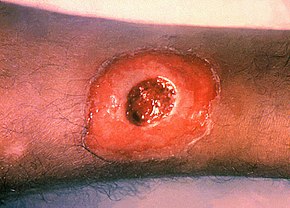
Diphtheria (Greek διφθερα (diphthera) — “pair of leather scrolls”), is an upper respiratory tract illness characterized by sore throat, low-grade fever, and an adherent membrane (a pseudomembrane) on the tonsils, pharynx, and/or nasal cavity. A milder form of diphtheria can be restricted to the skin. It is caused by Corynebacterium diphtheriae, a facultatively anaerobic Gram-positive bacterium.
Once quite common, diphtheria has largely been eradicated in developed nations through wide-spread vaccination. In the United States for instance, between 1980 and 2004 there have been 57 reported cases of diphtheria (and only five cases since 2000) as the DPT (Diphtheria-Pertussis-Tetanus) vaccine is given to all school children. Boosters of the vaccine are recommended for adults since the benefits of the vaccine decrease with age; they are particularly recommended for those traveling to areas where the disease has not been eradicated.
Diphtheria was named in 1826 by French physician Pierre Bretonneau. The name alludes to the leathery, sheath-like membrane that grows on the tonsils, throat, and in the nose. The pronunciation /ˌdipˈθiɹˌi.ə/ was originally considered incorrect, but has become the most common way of saying the word, and is accepted as a correct form. While many writers today use the spelling "diptheria" which fits the modern pronunciation, this spelling is rarely found in dictionaries.
Diphtheria was once a dreaded disease, with frequent large-scale outbreaks. A diphtheria epidemic in the New England colonies between 1735 and 1740 was said to have killed as many as 80% of the children under 10 years of age in some towns.

In the 1920s there were an estimated 100,000 to 200,000 cases of diphtheria per year in the United States, causing 13,000 to 15,000 deaths. Children represented a large majority of these cases and fatalities. One of the most famous outbreaks of diphtheria was in Nome, Alaska; the trip made to get the antitoxin is now celebrated by the Iditarod Trail Sled Dog Race.
One of the first effective treatments for diphtheria was discovered in the 1880s by U.S. physician Joseph O'Dwyer (1841-1898). O'Dwyer developed tubes that were inserted into the throat, and prevented victims from suffocating due to the membrane sheath that grows over and obstructs airways. In the 1890s, the German physician Emil von Behring developed an antitoxin that did not kill the bacteria, but neutralized the toxic poisons that the bacteria releases into the body. von Behring was awarded the first Nobel Prize in Medicine for his role in the discovery, and development of a serum therapy for diphtheria. Americans William H. Park and Anna Wessels Williams; and Pasteur Institute scientists Emile Roux and Auguste Chaillou also independently developed diphtheria antitoxin in the 1890s. The first successful vaccine for diphtheria was developed in 1923. However, antibiotics against diphtheria were not available until the discovery and development of sulfa drugs following World War II.
Signs and Symptoms
The respiratory form has an onset of disease, which is usually gradual. Symptoms include fatigue, fever, a mild sore throat and problems in swallowing. Children infected have symptoms that include nausea, vomiting, chills, and a high fever, although some do not show symptoms until the infection has progressed further. In 10% of cases, patients experience neck swelling. These cases are associated with a higher risk of death.
In addition to symptoms at the site of infection (sore throat), the patient may experience more generalized symptoms, such as listlessness, pallor, and fast heart rate. These symptoms are caused by the toxin released by the bacterium. Low blood pressure may develop in these patients. The toxin, or poison caused by the bacteria can also lead to a thick coating in the nose, throat, or airway. This coating is usually fuzzy gray or black and is responsible for breathing problems and difficulty in swallowing. Longer-term effects of the diphtheria toxin include cardiomyopathy and peripheral neuropathy (sensory type).
The cutaneous form of diphtheria is often a secondary infection of a preexisting skin disease. This form of diphtheria causes sores on the skin that may be painful, red, and swollen, and may also have patches of a sticky, gray material.
Causative Agent
Mode of Transmission
Diphtheria is a highly contagious disease spread by direct physical contact or breathing the aerosolized secretions of infected individuals.
Diagnosis
Diagnosis is usually made based on the clinical presentation since it is imperative to begin presumptive therapy quickly.
Culture of the lesion is done to confirm the diagnosis. It is critical to take a swab of the pharyngeal area, especially any discolored areas, ulcerations, and tonsillar crypts. Culture medium containing tellurite is preferred because it provides a selective advantage for the growth of this organism. A blood agar plate is also inoculated for the detection of hemolytic streptococcus. If diphtheria bacilli are isolated, they must be tested for toxin production.
Gram stain and Kenyon stain of material from the membrane itself can be helpful when trying to confirm the clinical diagnosis. The Gram stain may show multiple club-shaped forms which look like Chinese characters. Other Corynebacterium species (“diphtheroids”) that can normally inhabit the throat may confuse the interpretation of direct stain. However, treatment should be started if clinical diphtheria is suggested, even in the absence of a diagnostic Gram stain.
Incubation Period
Pathogenesis / Pathophysiology
Diphtheria organisms usually remain in the superficial layers of skin lesions or respiratory mucosa, inducing local inflammatory reaction. The organism's major virulence lies in its ability to produce the potent 62-kd polypeptide exotoxin, which inhibits protein synthesis and causes local tissue necrosis.
Toxigenic strains of C. diphtheriae carry the tox structural gene found in lysogenic corynebacteriophages beta-tox+, gamma-tox+, and omega-tox+.
Highly toxic strains have 2 or 3 tox+ genes inserted into the genome. Expression of the gene is regulated by the bacterial host and is iron dependent. In the presence of low concentrations of iron, the gene regulator is inhibited, resulting in increased toxin production. Toxin is excreted from the bacterial cell and undergoes cleavage to form 2 chains, A and B, which are held together by an interchain disulfide bond between cysteine residues at positions 186 and 201. As toxin concentrations increase, the toxic effects extend beyond the local area because of distribution of the toxin by the circulation. Diphtheriae toxin does not have a specific target organ, but myocardium and peripheral nerves are most affected.
Within the first few days of respiratory tract infection, a dense necrotic coagulum of organisms, epithelial cells, fibrin, leukocytes, and erythrocytes forms, advances, and becomes a gray-brown adherent pseudomembrane. Removal is difficult and reveals a bleeding edematous submucosa. Paralysis of the palate and hypopharynx is an early local effect of the toxin. Toxin absorption can lead to necrosis of kidney tubules, thrombocytopenia, cardiomyopathy, and demyelination of nerves. Since cardiomyopathy and demyelination of nerves can occur 2-10 weeks after mucocutaneous infection, the pathophysiologic mechanism may be immunologically mediated in some patients.
In the classic description of diphtheria, the primary focus of infection is the tonsils or pharynx in more then 90% of patients; the nose and larynx are the next two most common sites. After an average incubation period of 2-5 days, local signs and symptoms of inflammation develop. Fever is rarely higher than 39°C.
Prevention
The only effective control measure against diphtheria is universal immunization with diphtheria toxoid throughout life to provide constant protective antitoxin levels and to reduce indigenous C. diphtheriae. Although immunization does not preclude subsequent respiratory or cutaneous carriage of toxigenic C. diphtheriae, it decreases local tissue spread, prevents toxic complications, diminishes transmission of the organism, and provides herd immunity when at least 70-80% of a population is immunized. Serum antitoxin concentration of 0.01 IU/mL is conventionally accepted as the minimum protective level, and 0.1 IU/mL provides a definitely protective level.
Nursing Interventions
- Maintain room temperature and advise the client to wear thin clothes that absorb sweat easily.
- Encourage fluid intake, and administer antipyretics as ordered.
- Assess for hoarseness, stridor, shortness of breath, and cyanosis.
- Keep patient on strict bed rest, strict isolation.
- Room should have adequate means of ventilation.
- Provide cleansing throat gargle as ordered.
- Monitor calorie intake and quality of food consumption; give foods that stimulate the appetite, and measure body weight daily.
- Observe for respiratory obstruction (tracheotomy).
- Use suctioning as needed (tracheotomy).
- O2 therapy as ordered.
- Administer Antitoxin against toxin (as ordered).
- Administer toxoid to immunized contact (as ordered).
- Administer Broad spectrum antibiotic against diphtheria bacilli (as ordered).
- Provide Health teaching on proper hygiene and universal precaution.
- Monitor Vital signs.
- Provide oral care as the mouth, teeth and lips demand careful attention.
Treatment
The disease may remain manageable, but in more severe cases lymph nodes in the neck may swell, and breathing and swallowing will be more difficult. People in this stage should seek immediate medical attention, as obstruction in the throat may require intubation or a tracheotomy. In addition, an increase in heart rate may cause cardiac arrest. Diphtheria can also cause paralysis in the eye, neck, throat, or respiratory muscles. Patients with severe cases will be put in a hospital intensive care unit (ICU) and be given a diphtheria anti-toxin. Since antitoxin does not neutralize toxin that is already bound to tissues, delaying its administration is associated with an increase in mortality risk. Therefore, the decision to administer diphtheria antitoxin is based on clinical diagnosis, and should not await laboratory confirmation.
Antibiotics have not been demonstrated to affect healing of local infection in diphtheria patients treated with antitoxin. Antibiotics are used in patients or carriers to eradicate C. diphtheriae and prevent its transmission to others. The CDC recommends either:
- Erythromycin (orally or by injection) for 14 days (40 mg/kg per day with a maximum of 2 g/d), or
- Procaine penicillin G given intramuscularly for 14 days (300,000 U/d for patients weighing <10 kg and 600,000 U/d for those weighing >10 kg). Patients with allergies to penicillin G or erythromycin can use rifampin or clindamycin.
Complications
Demyelination of nervous tissue is seen in all fatal cases of diphtheria.
Frank paralysis occurs in 10-20% of patients and most often involves the muscles of the palate and the hypopharynx, beginning as early as the first 10 days of illness.
Difficulty swallowing and nasal speech often are the first indications of neurologic impairment.
Involvement of other cranial nerves, which may be delayed until as late as 7 weeks after infection, results in oculomotor paralysis and blurred vision. Diffuse, usually bilateral, motor function deficits resulting from involvement of the anterior horn cells of the spinal cord may be seen as late as 3 months after initial disease, with progression of weakness either from proximal-to-distal regions or, more commonly, from distal-to-proximal regions.
Involvement of the phrenic nerve may cause diaphragmatic paralysis at any time between the first and seventh weeks of illness.
Elevation of CSF protein levels can be seen and may lead to an erroneous diagnosis of Guillain-Barré syndrome.
Recovery from neurologic damage usually is complete in patients who survive.
Cardiac complications may arise during the first 10 days of illness or may be delayed until 2-3 weeks after onset, when pharyngeal disease is subsiding. Cardiac involvement is thought to be responsible for 50-60% of deaths associated with diphtheria.
The first sign of toxin-induced myocardiopathy is tachycardia disproportionate to the degree of fever.
A variety of dysrhythmias, including first-, second-, or third-degree heart block; atrioventricular dissociation; and ventricular tachycardia can develop, and congestive heart failure may be a consequence of myocardial inflammation.
Echocardiogram may demonstrate dilated or hypertrophic cardiomyopathy.
In patients who survive, cardiac muscle regeneration and interstitial fibrosis lead to recovery of normal cardiac function, unless toxic damage has led to a permanent arrhythmia.
Airway obstruction by the diphtheritic membrane and peripharyngeal edema combine to pose a risk of death in patients with diphtheria.
