Tachycardia, also known as tachyarrhythmia, is a medical term for a heart rate that exceeds 100 beats a minute. Tachycardia may be normal if it is due to exercise or as a response to stress. But Tachycardia is considered abnormal if it is caused by an underlying medical conditions such as electrical problems within the heart, and ischemia. Many types of irregular heart rhythms (arrhythmias) can cause tachycardia.
The term comes from Greek ταχύς — tachys, which means "quick, rapid" and καρδία — kardia, which means "heart".
Tachycardia may not cause any symptoms or complications, but if left untreated, some forms of tachycardia can lead to serious health problems, including stroke, heart failure, or sudden cardiac arrest.
Types
Various forms of tachycardia exist, each with distinct characteristics.
- Sinus tachycardia: This type is triggered by factors like physical exertion or stress.
- Atrial Fibrillation (A-fib): The most prevalent form, characterized by disordered electrical signals in the heart's upper chambers (atria), inducing rapid heartbeats. While some occurrences might be temporary, others persist until treated.
- Atrial Flutter: Similar to A-fib but with more organized heartbeats. Episodes may resolve spontaneously or necessitate intervention. Individuals experiencing atrial flutter often concurrently suffer from atrial fibrillation.
- Ventricular Tachycardia: Originating in the lower heart chambers (ventricles), this arrhythmia impedes proper ventricular filling and contraction, affecting blood pumping. Brief episodes might be harmless, but prolonged instances can be life-threatening.
- Supraventricular Tachycardia (SVT): Encompassing arrhythmias originating above the ventricles. SVT triggers abrupt, pounding heartbeats (palpitations) that start and stop suddenly.
- Ventricular Fibrillation: Rapid, disorganized electrical impulses cause the ventricles to quiver instead of contracting in a coordinated way. This severe condition can lead to death if normal heart rhythm isn't restored promptly. Most individuals experiencing ventricular fibrillation either have an underlying heart ailment or have encountered severe trauma, such as being struck by lightning.
Signs
- Sensation of a racing, pounding heartbeat or flopping in the chest (palpitations)
- Chest pain
- Fainting (syncope)
- Lightheadedness
- Rapid pulse rate
- Shortness of breath
Diagnosis
1. Medical history
- Previous episodes of tachycardia
- Any existing medical conditions
- Current medications
- Family history of heart problems
- Lifestyle habits (e.g., smoking, alcohol consumption, caffeine intake)
2. Physical examination: Done by assessing the heart rate, rhythm, blood pressure, and other vital signs. Also by listening to heart sounds for any abnormalities.
3. Tests:
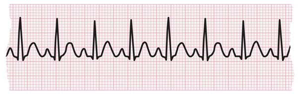
- Electrocardiogram (ECG or EKG): This test records the electrical activity of the heart and can help identify the type of tachycardia.
- Holter monitor: This device is worn for 24-48 hours and continuously records the heart rhythm.
- Event recorder: This small device is worn for a longer period and automatically records the heart rhythm when the patient experiences symptoms.
- Echocardiogram: This test uses ultrasound waves to produce images of the heart and assess its structure and function.
- Blood tests: These tests can check for various conditions that can cause tachycardia, such as anemia, thyroid problems, or electrolyte imbalances.
Based on the information gathered from the medical history, physical examination, and tests, a healthcare professional can diagnose the specific type of tachycardia and recommend the appropriate treatment.
Nursing Interventions
1. Monitoring:
- Monitor vital signs, including heart rate, rhythm, blood pressure, and oxygen saturation, frequently.
- Assess for any associated symptoms like chest pain, shortness of breath, dizziness, or fainting.
- Monitor the patient's response to treatment.
- Document all findings and interventions accurately and promptly.
2. Promoting Vagal Maneuvers:
- Encourage the patient to perform vagal maneuvers, which can stimulate the vagus nerve and help slow down the heart rate. Examples of vagal maneuvers include:
- Valsalva maneuver: The patient holds their breath and bears down like they are having a bowel movement.
- Carotid massage: Gently massaging the carotid artery in the neck (only to be performed by a healthcare professional).
- Cold face immersion: Immersing the face in cold water.
3. Managing Underlying Cause:
- Administer medications as prescribed by the physician to slow down the heart rate and address the underlying cause of the tachycardia. Common medications used for tachycardia include beta-blockers, calcium channel blockers, and antiarrhythmic drugs.
- Provide oxygen therapy if the patient is hypoxic.
- Maintain fluid and electrolyte balance to ensure optimal heart function.
- Manage any pain or discomfort the patient may be experiencing.
- Educate the patient about their condition and its treatment plan.
- Offer emotional support and reassurance to the patient.
- Collaborate with the physician and other healthcare professionals to provide the best possible care for the patient.