
Bladder cancer refers to any of several types of malignant growths of the tissues of the urinary bladder (the organ that stores urine). It is a disease in which abnormal cells multiply without control in the bladder. The most common type of bladder cancer begins in cells lining the inside of the bladder and is called transitional cell carcinoma (sometimes urothelial cell carcinoma). Other types include squamous cell carcinoma (cancer that begins in thin, flat cells) and adenocarcinoma (cancer that begins in cells that make and release mucus and other fluids). The cells that form squamous cell carcinoma and adenocarcinoma develop in the inner lining of the bladder as a result of chronic irritation and inflammation.
Signs and Symptoms
Bladder cancer characteristically causes blood in the urine; this may be visible to the naked eye (frank hematuria) or detectable only by microscope (microscopic hematuria). Other possible symptoms include pain during urination, frequent urination (Polyuria) or feeling the need to urinate without results (anuria). These signs and symptoms are not specific to bladder cancer, and are also caused by non-cancerous conditions, including prostate infections and cystitis. Kidney cancer can also cause hematuria.
Other signs and symptoms include back or abdominal pain, and loss of appetite and weight.
Causes
Tobacco smoking is the main known cause of urinary bladder cancer: in most populations, smoking causes over half of bladder cancer cases in men and a sizeable proportion in women. There is a linear relationship between smoking and risk, and quitting smoking reduces the risk. In a 10-year study involving almost 48,000 men, researchers found that men who drank 1.5L of water a day had a significantly reduced incidence of bladder cancer when compared with men who drank less than 240mL (around 1 cup) per day. The authors proposed that bladder cancer might partly be caused by the bladder directly contacting carcinogens that are excreted in urine. Thirty percent of bladder tumors probably result from occupational exposure in the workplace to carcinogens such as benzidine. 2-Naphthylamine, which is found in cigarette smoke, has also been shown to increase bladder cancer risk. Occupations at risk are metal industry workers, rubber industry workers, workers in the textile industry, and people who work in printing. Some studies also suggest that auto mechanics have an elevated risk of bladder cancer due to their frequent exposure to hydrocarbons and petroleum-based chemicals. Hairdressers are thought to be at risk as well because of their frequent exposure to permanent hair dyes.
Diagnosis
Radiology
An Intravenous pyelogram (IVP) is a conventional x-ray test using dye to examine the pelves of the kidneys (where urine collects within the kidneys), ureters, and bladder. This x-ray allows visualization of the upper and lower urinary tract to determine the presence of any abnormality.
Computed Tomography (CT) scanning
It is essentially a detailed X-ray of the body. CT shows cross-sections of the body and allows your doctor to see details of the anatomy that would not be seen on regular x-ray.
Magnetic Resonance Imaging (MRI)
It is more sensitive than CT scanning. CT and MRI have the added benefit of detecting enlarged lymph nodes near the tumors, which can suggest that a cancer has spread (metastasized) to the lymph nodes.
Cystoscopy
Cystoscopy is performed by the urologist. It evaluates the bladder by direct visual examination with a specialized instrument called a cystoscope, which is placed in the bladder via the urethra during the examination. The purpose of routine outpatient cystoscopy is to evaluate the lining of the lower urinary tract. If abnormalities such as tumors, stones, or patches of abnormal appearing tissue are discovered during cystoscopy, a biopsy may be taken at that time.
Pathology
The diagnosis of bladder cancer is based on examining cells from the bladder, either from a urine specimen or biopsy of the bladder. Only a pathologist can diagnose if a bladder cancer is present and the type of bladder cancer, by looking at the bladder tissue. The correct diagnosis is critical, as appropriate treatment of bladder cancer is dependent upon the type of cancer seen. The pathology of the bladder is complex and therefore a second opinion is often advisable and can have a major impact in therapy.
The diagnosis can sometimes be made by examining urine cytology. A cytopathologist looks at individual cells from the urine, which are spread into a thin layer onto glass microscopic slides. These procedures have the benefit of not requiring an operation or general anesthesia.
Biopsy of the bladder, performed through the cystoscope, is the more common means of diagnosing these tumors. The pathologist will examine a small sample (a biopsy) of the bladder tissue under a microscope. The pathologist identifies whether the tumor is benign or malignant and the type of tumor. This is essential because tumors of different types behave very differently and require different treatment regimens.
Pathogenesis / Pathophysiology
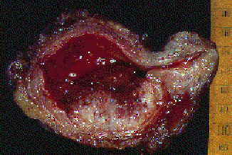
Almost all bladder cancers are epithelial in origin. The urothelium consists of a 3- to 7-cell mucosal layer within the muscular bladder. Of these urothelial tumors, more than 90% are transitional cell carcinomas. However, up to 5% of bladder cancers are squamous cell in origin, and 2% are adenocarcinomas. Nonurothelial primary bladder tumors are extremely rare and may include small cell carcinoma, carcinosarcoma, primary lymphoma, and sarcoma.
Bladder cancer is often described as a polyclonal field change defect with frequent recurrences due to a heightened potential for malignant transformation. However, bladder cancer has also been described as a problem with implantation and migration from a previously affected site.
The World Health Organization classifies bladder cancers as low grade (grade 1 and 2) or high grade (grade 3). Tumors are also classified by growth patterns: papillary (70%), sessile or mixed (20%), and nodular (10%). Carcinoma in situ (CIS) is a flat, noninvasive, high-grade urothelial carcinoma. The most significant prognostic factors for bladder cancer are grade, depth of invasion, and the presence of CIS.
Upon presentation, 55-60% of patients have low-grade superficial disease, which is usually treated conservatively with transurethral resection and periodic cystoscopy. Forty to forty-five percent of patients have high-grade disease, of which 50% is muscle invasive and is typically treated with radical cystectomy.
Less than 5% of bladder cancers in the United States are squamous cell carcinomas (SCCs). However, worldwide, SCC is the most common form, accounting for 75% of bladder cancer in underdeveloped nations. In the United States, SCC is associated with persistent inflammation from long-term indwelling Foley catheters and bladder stones. In underdeveloped nations, SCC is associated with bladder infection by Schistosoma haematobium.
Adenocarcinomas account for less than 2% of primary bladder tumors. These tumors are observed most commonly in exstrophic bladders and respond poorly to radiation and chemotherapy. Radical cystectomy is the treatment of choice.
Small cell carcinomas are aggressive tumors associated with a poor prognosis and are thought to arise from neuroendocrine stem cells.
Carcinosarcomas are highly malignant tumors that contain both mesenchymal and epithelial elements.
Primary bladder lymphomas arise in the submucosa of the bladder and are treated with radiation therapy.
Leiomyosarcoma is the most common sarcoma of the bladder.
Rhabdomyosarcomas most commonly occur in children and carry a poor prognosis.
Race
Bladder cancer is more common in whites than in blacks; however, blacks have a worse prognosis than whites.
Sex
The male-to-female ratio is 3:1. Women generally have a worse prognosis than men.
Age
The median age at diagnosis is 68 years, and the incidence increases with age.
Prevention
Bladder cancer cannot be prevented, but it is possible to reduce some of the risk factors that develops it.
- Cigarette smokers are much more likely to develop bladder cancer than nonsmokers.
- Avoid exposure to industrial chemicals, such as benzene substances and arylamines. Occupational exposure from working with dyes, rubbers, textiles, paints, leathers, and chemicals increases the risk of developing bladder cancer.
- Avoid exposure to arsenic. Test drinking water, and/or drink bottled water if suspected of contamination.
- Eating healthy diet.
- Eating low-fat, low-cholesterol diet that includes plenty of fruits and vegetables.
- Avoid dehydration. Increase fluid intake, particularly water. Water dilutes cancer-causing chemicals.
Nursing Interventions
Preoperative Care:
- Assessment and Education: Conduct a thorough assessment of the patient's medical history, current symptoms, and overall health status. Provide comprehensive education about bladder cancer, its diagnosis, treatment options, and potential side effects.
- Emotional Support: Offer emotional support and counseling to help patients cope with the diagnosis and prepare for treatment. Address their fears, concerns, and anxiety related to bladder cancer and its impact on their lives.
- Stoma Care Education: If stoma creation is planned, provide detailed education about stoma care, including stoma management techniques, appliance selection, skin care, and troubleshooting potential problems.
Postoperative Care:
- Pain Management: Assess and manage pain effectively using appropriate medications and non-pharmacological interventions, such as relaxation techniques and positioning.
- Wound Care: Provide meticulous wound care to promote healing and prevent complications. Monitor for signs of infection and address any wound-related issues promptly.
- Fluid Management: Ensure adequate fluid intake to maintain hydration and prevent urinary tract infections. Monitor fluid balance and electrolyte levels closely.
- Bladder Irrigation: If bladder irrigation is prescribed, provide proper instruction and support to the patient regarding the irrigation procedure, catheter insertion and removal, and the importance of maintaining aseptic technique.
- Catheter Care: If a urinary catheter is present, provide thorough instruction on catheter care, including cleaning, maintenance, and signs of potential complications.
- Urinary Diversion Management: If a urinary diversion is present, such as an ileal conduit or urostomy, provide comprehensive education and support regarding the management of the diversion method, including appliance selection, drainage techniques, and skin care.
- Nutritional Counseling: Provide nutritional counseling to ensure the patient maintains a healthy diet and adequate hydration to support healing and overall well-being.
- Psychosocial Support: Continue to offer emotional support and counseling to help patients adjust to their condition, cope with treatment-related challenges, and maintain a positive outlook.
- Follow-up Care: Schedule regular follow-up appointments to monitor the patient's progress, assess for recurrence, provide ongoing education, and address any new concerns or symptoms.
Treatment
The treatment of bladder cancer depends on how deep the tumor invades into the bladder wall. Superficial tumors (those not entering the muscle layer) can be "shaved off" using an electrocautery device attached to a cystoscope. Immunotherapy in the form of BCG instillation is also used to treat and prevent the recurrence of superficial tumors. BCG immunotherapy is effective in up to 2/3 of the cases at this stage. Instillations of chemotherapy, such as valrubicin (Valstar) into the bladder can also be used to treat BCG-refractory CIS disease when cystectomy is not an option.
Untreated, superficial tumors may gradually begin to infiltrate the muscular wall of the bladder. Tumors that infiltrate the bladder require more radical surgery where part or all of the bladder is removed (a cystectomy) and the urinary stream is diverted. In some cases, skilled surgeons can create a substitute bladder (a neobladder) from a segment of intestinal tissue, but this largely depends upon patient preference, age of patient, renal function, and the site of the disease.
A combination of radiation and chemotherapy can also be used to treat invasive disease. It has not yet been determined how the effectiveness of this form of treatment compares to that of radical ablative surgery.
The hemocyanin found in Concholepas concholepas blood has immunotherapeutic effects against bladder and prostate cancer. In a research made in 2006, mice were primed with C. concholepas before implantation of bladder tumor (MBT-2) cells. Mice treated with C. concholepas showed a significant antitumor effect as well. The effects included prolonged survival, decreased tumor growth and incidence and lack of toxic effects.
Complications
Bladder cancers may spread into the nearby organs. They may also travel through the pelvic lymph nodes and spread to the liver, lungs, and bones. Additional complications of bladder cancer include:
- Anemia
- Swelling of the ureters (hydronephrosis)
- Urethral stricture
- Urinary incontinence
Prognosis
Patients are closely monitored to see whether the disease gets worse, regardless of which kind of treatment they received. Monitoring may include:
- Bone scan and/or CT scan to check for cancer spread
- Checking for other signs of disease progression, such as fatigue, weight loss, increased pain, decreased bowel and bladder function, and weakness
- Complete blood count (CBC) to monitor for anemia
- Cystoscope evaluations every 3 to 6 months after treatment
- Urine cytology evaluations (for people whose bladder has not been removed)
How well a patient does depends on the initial stage and response to treatment of the bladder cancer. The outlook for stage 0 or I cancers is fairly good. Although the risk of the cancer returning is high, most bladder cancers that return can be surgically removed and cured.
The cure rates for people with stage III tumors are less than 50%. Patients with stage IV bladder cancer are rarely cured.